• +91 9718599220
  • +91 9968569870
  • info@cardiolyfe.com
  • 8/26, West Patel Nagar, Delhi-08
|| Booking Appointment

Consult your heart speciality now

|| Expert Team

Adept Cardiac Service Provider Team

Dr. Sudhanshu Sekhar Parida

Dr. Sudhanshu Sekhar Parida

Founder CEO

Dr.Sudhansu Sekhar Parida is one of the renowned Interventional Cardiologists in New Delhi. He is currently working as a Senior Consultant, (Interventional Cardiology) at MAX Super Speciality Hospital, Shalimar Bagh. He is highly qualified, having done his Post Graduation from most reputed institute of India - AIIMS Delhi and Super-speciality from G B Pant Hospital Delhi.

He has rich experience in Interventional Cardiology procedures and has performed more than 20000 angiographies & angioplasties as a primary operator. He is an expert in Transradial Angiography, i.e., angiography via the radial (wrist) route, which is considered to be safer and patient-friendly. He is well versed in all state-of-the-art technologies that include interventions like Rota Ablation, & Atherectomy, Fractional Flow Reserve (FFR), and intracoronary imaging like Intra Vascular Ultrasound (IVUS) and Optical Coherence Tomography (OCT). He also has experience in device closure for various types of congenital heart disease like atrial septal defects (ASD), ventricular septal defects, (VSD) & patent ductus arteriosus (PDA). He has also done numerous cases of ballooning of blocked valves like BMV, AVBD, PBVD. He also has immense experience and expertise with permanent pacemaker implantation, AICD, CRT-P, and CRT-D. Other interventions in which he has experience like peripheral, carotid, and renal angioplasties, IVC filter, PERMACATH insertion, etc.

Cardiologist
|| Services

We give the best
Cardiac services

Electrocardiogram (ECG)

An electrocardiogram (ECG) is one of the simplest and fastest tests, used to evaluate the heart. Electrodes are placed at certain spots on the chest, arms, and legs. The electrodes are connected to an ECG machine by lead wires. the electrical activity of the heart is then measured, interpreted, and printed out it is used to look for the cause of chest pain and also to evaluate problems that may be heart-related like to identify irregular heartbeats.

Echocardiograhy (ECHO)

An echocardiogram is an ultrasound of the heart that uses high-frequency sound waves to capture pictures of the heart’s valves and chambers, allowing a cardiologist to evaluate the pumping action of the heart. Echo is often combined with Doppler ultrasound and color Doppler to evaluate blood flow across the heart’s valves. An echocardiography test is used to assess the overall function of your heart and determine the presence of many types of heart disease. Tests do not require fasting. During the test, the healthcare provider will explain the procedure in detail. A cardiologist will place electrodes on your chest that connect to an electrocardiograph (ECG) monitor to chart your heart’s electrical activity. You may be asked to change positions several times during the exam so the cardiologist can take pictures of different areas of the heart. You should feel no major discomfort. You may feel a coolness on your skin from the gel on the transducer, and a slight pressure of the transducer on your chest

Treadmill Test (TMT)

The tread mill stress test records the heart’s electrical activity, including rate and rhythm, during exercise, most often for the diagnosis of coronary artery disease. Exercise places stress on the heart, and performing a treadmill stress test can bring out abnormalities caused by partial blockages in the coronary arteries – abnormalities that may not be apparent at rest. During the test technician will place electrodes on your chest, You will be asked to perform a “graded” exercise test on a treadmill. The exercise protocol that you will follow will be determined by the cardiologist supervising your test but will begin at a relatively easy level and become progressively more difficult with each subsequent stage Your blood pressure, heart rate, and ECG will be recorded at frequent intervals at rest and during exercise and after exercise. The physician or technologist may stop the test at any time for medical reasons, and you may ask to stop the test at any time due to fatigue or discomfort.

Stress ECHO

A “stress echo” is a combination of an echocardiography and exercise test. It compares how your heart muscle is pumping before and after exercise. This test takes about 40 minutes. During the procedure, the cardiologist will take images of your heart whilst at rest. A water-soluble gel is used as a contact medium between the probe and your chest wall to improve image quality. You will feel pressure on your chest where the sonographer is imaging. Once the resting images have been taken you will be connected to an ECG machine used to perform a standard treadmill exercise test. Your cardiologist will be present throughout this part of the test and your blood pressure, heart rate and symptoms will be constantly monitored. The treadmill will increase in speed and slope. It will be stopped when you cannot walk/run any further when you have reached an appropriate target or at the cardiologist’s discretion. At the end of the exercise test, the sonographer will take more images of your heart before your heart rate slows down. The cardiologist will compare the resting images to those taken at maximum exertion.

Dobutamine Stress Echo (DSE)

A “stress echo” is a combination of an Echocardiography and exercise test. It compares how your heart muscle is pumping before and after exercise. This test takes approximately 40 minutes. A Dobutamine stress echo ( DSE )is performed when your doctor feels that it is inappropriate for you to exercise on the treadmill. Dobutamine is a medication administered to place your heart under a similar amount of strain as would be achieved with exercise. This information can assist your cardiologist in assessing the state of your heart muscle and the arteries feeding that muscle During the procedure, you will be weighed to calculate the appropriate dose of Dobutamine. Next, a small intravenous line will be inserted by a nurse. You will also be connected to an ECG machine for the duration of the test. The cardiac sonographer will begin by taking images of your heart whilst at rest. A water-soluble gel is used as a contact medium between the probe and your chest wall to improve image quality. You will feel pressure on your chest where the sonographer is imaging. You will then be given an intravenous infusion of Dobutamine and the medication will be increased every 2 minutes. At certain intervals, more images of your heart will be acquired. You can expect to feel your heart pounding and possibly even some tingling sensations in your face as a result of the medication. Your cardiologist will be present throughout this part of the test and your blood pressure, heart rate and symptoms will be constantly monitored. When your heart rate has increased sufficiently or at the cardiologist’s discretion, the Dobutamine infusion will be ceased. The cardiologist will compare the resting images of your heart with those taken at each interval.

Holter Test

A Holter monitor is a small, medical device that measures your heart’s activity, such as rate and rhythm for 24 hours. This device is connected to the electrodes and electrical leads, and even newer devices use only a single patch to detect hearts activity, which is more convenient for the patients. Holter monitor testing is also sometimes called Ambulatory Electrocardiography. We ask that you keep a diary while you wear the monitor, detailing your activities and your symptoms, which may include palpitations, rapid heartbeat, dizziness, or lightheadedness. Make sure you note the time, and what activities you were doing when your symptoms occurred so that we are able to see the correlation between the two. The electrical impulses are continuously recorded and stored in the Holter monitor. When you return the Holter monitor to us, our team will analyze the recording and look for any rhythm abnormalities. When the full report is available, a doctor will discuss the results with you

External Loop Recorder (ELR)

An External Loop Recorder (ELR) is a small, medical device that measures your heart’s activity, such as rate and rhythm for an extended period of time. This device is a compact lightweight patch to detect heart activity monitoring from 24 hours to 14 days. It is usually water and dust-proof. It is used for various rhythm abnormalities which cant be picked up by Holter monitoring. The ELR system, primarily consists of a patch, like a small device put on the chest. the patch records arrhythmia episodes, which are stored in the device. the device is dust and waterproof allowing all indoor and outdoor activity. you’re encouraged to participate in your normal activities during the ELR test. After test completion, our team will analyze the recording from stored data in the device and look for any rhythm abnormalities. When the full report is available, a doctor will discuss the results with you

Ambulatory Blood Pressure Monitoring (ABPM)

Twenty-four-hour ambulatory blood pressure monitoring (ABPM) is a way of measuring and managing high blood pressure (hypertension). It allows your blood pressure (BP) readings to be recorded over a 24-hour period, whether you’re awake or asleep. In most cases, readings are taken every 20 to 30 minutes during the day and every hour at night. Your heart rate can be measured at the same time. During the procedure, BP readings are taken continually over a 24-hour period. You’ll wear a device that is about the same size as a portable radio. The device is attached to a belt or strap worn on your body. You’ll wear a bp cuff that is attached to the device around your upper arm. The cuff inflates at certain intervals throughout the day and night. After 24 hours, you can remove the device and bp cuff. These multiple readings are averaged over the 24-hour period. Changes in BP and heart rate, the BP distribution pattern, and other statistics are calculated.

Coronary Angiogram (CAG)

A coronary angiogram ( CAG ) is a procedure that uses X-ray imaging to see your heart's blood vessels. The test is generally done to see if there's a blockage in arteries, that supply blood flow to the heart. During a coronary angiogram, a type of dye that's visible by an X-ray machine is injected into the blood vessels of your heart. The X-ray machine rapidly takes a series of images (angiograms), offering a look at your blood vessels. If necessary, An angiogram can help your doctor see if you need treatment such as angioplasty(stenting) or coronary artery bypass surgery (CABG) During CAG, a long, narrow tube called a catheter is inserted through a plastic introducer sheath (a short, hollow tube that is inserted into a blood vessel in your arm or leg. The catheter is guided through the blood vessel to the coronary arteries with the aid of a special X-ray machine. Contrast material is injected through the catheter and X-ray movies are created as the contrast material moves through the heart’s blood vessels. The digital photographs of the contrast material are used to identify the site of the narrowing or blockage in the coronary artery. The patient will be awake and conscious during the entire procedure.

Coronary Angioplasty

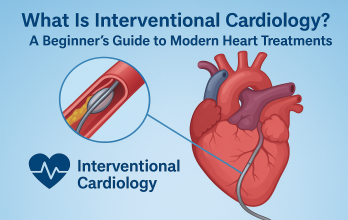
Coronary angioplasty is a non-surgical procedure used to open narrowed coronary arteries to improve blood flow to the heart. This can be performed during a coronary angiogram when a blockage is identified, or it may be scheduled after a CAG has confirmed the presence of a blockage. Angioplasty can be balloon Angioplasty Or stenting. In most cases, balloon angioplasty is performed in combination with the stenting procedure. A stent is a small, metal mesh tube that acts as a scaffold to provide support inside the coronary artery. It can be both Bare-metal Stent (BMS) or Drug-Eluting Stent (DES). Once in place, the balloon is inflated and the stent expands to the size of the artery and holds it open. The balloon is deflated and removed, and the stent stays in place permanently. The patient will be awake and conscious during the entire procedure

Permanent Pacemaker Implantation (PPI)

A pacemaker is a device that sends small electrical impulses to the heart muscle to maintain a suitable heart rate. it can be either a Single Chamber Pacemaker or Dual Chamber Pacemaker. Your doctor will decide what type of pacemaker you need based on your heart condition. Your doctor also determines the minimum rate to set your pacemaker. When your heart rate drops below the set rate, the pacemaker generates an impulse that passes through the lead to the heart muscle. This causes the heart muscle to contract, creating a heartbeat The procedure takes about an hour. During this time you will be given some light sedation to help you relax and feel comfortable. You will be awake. An area below the collar bone will be numbed with local anesthetic and the cardiologist will make a small cut (approx 5-8cms) to insert the pacemaker. The leads are then guided through a vein into the heart and connected to the device. The skin is then sewn together and a small dressing applied

Implantable cardioverter defibrillator (ICD)

An implantable cardioverter-defibrillator (ICD) is an electronic device that constantly monitors your heart rhythm. When it detects a very fast, abnormal heart rhythm, it delivers energy to the heart muscle. This causes the heart to beat in a normal rhythm again. It can be either Single Chamber or Dual Chamber ICD. Ventricular tachycardia and ventricular fibrillation are two life-threatening heart rhythms that cause the heart to beat very fast. These conditions can be fatal if not treated immediately. Your doctor will recommend an ICD if you’ve had at least one episode of these heart rhythms or are at high risk of developing these types of heart rhythms. The procedure takes about an hour. During this time you will be given some light sedation to help you relax and feel comfortable. You will be awake. An area below the collar bone will be numbed with local anesthetic and the cardiologist will make a small cut (approx 5-8cms) to insert the AICD. The leads are then guided through a vein into the heart and connected to the device. The skin is then sewn together and a small dressing is applied.

|| Always Available

We are always ready To serve

We are committed to forming a true partnership with each patient as we strive to serve as guiding hands toward better health and a life lived to the fullest.

15+

Years Experience

5.0

Google Ratings

167+

Google Reviews


Photo Gallery  ||  Video Gallery

Some Of Our Happy Moments

|| Intervention

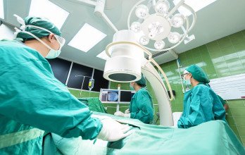
At Our Cathlab

|| Intervention

Rota Pro Demonstration

|| Miscellaaneous

Convocation

|| Intervention

AICD Implantation

|| Intervention

TAVR team

|| Miscellaaneous

COVID-19 & Heart Disease

|| Intervention

Complex PCI worshop

|| News

Get update About Cardiolyfe

blog

Living With Pulmonary Artery Hypertension

Pulmonary arterial hypertension (PAH) mainly affects the small pulmonary arterioles. It is a rare and progressive disease of the lungs.

blog

Impact of COVID infection and COVID vaccine in Heart disease patients

COVID-19 can cause worse outcomes in patients with pre-existing CVD risk-factors or in those with estabilished CVD.

blog

Effect Of Cold on Heart

Brief and prolonged exposure to cold both affect the cardiovascular system, and exercise in cold weather further increases stress on the heart and arteries.

|| Testimonial

What client say about us